ON THE CARE OF HEALTH.
by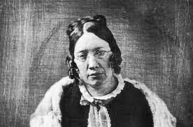 byCatharine Esther Beecher @catharinebeecher
Trailblazing American educator, advocated female education and kindergarten integration.
September 27th, 2023


Trailblazing American educator, advocated female education and kindergarten integration.


Trailblazing American educator, advocated female education and kindergarten integration.
About Author

Trailblazing American educator, advocated female education and kindergarten integration.
Comments